Thursday, April 18, 2013

Digestive System Part 1


Today's Medical Topic:  Digestive System Part I - If you aren't in my A&P II class you probably won't understand this.

What Are We Looking For:   The difference between the two liver models on our practical exam.


Here is one of the "livers" on our practical.  This one has more parts than the other ones so if you are asking yourself "Which one is more complicated?"  It is this one.  This is the only one that has a right and left hepatic duct.

To remember it correctly place the model of the liver in anatomical position and then rotate it up to see the vessels and what not.  The common hepatic duct on this model is a little confusing so let's clarify.


That little whitish part is actually the common hepatic duct on this model.  It is just a matter of how far up the cut is made.

Lets take a look at the other less complicated model:


You should position and rotate this one the same way and you can see how this one is different.  So basically if it has numbers you can see on it it is the less complicated model.  163 is the cystic duct, 162 is the common hepatic duct, and 165 is the bile duct.

So thats is all I got for tonight.  Enjoy.


Send questions or comments to dudaday@gmail.com

Disclaimer:  I am not a health care provider, any information presented in this blog should not be considered advice it is mearly an outlet to slake my curiosity.  You should always consult your primary medical provider for any concerns or illness.  Unlike Tylenol, I am not approved by the FDA or American Medical Association to treat or provide relief for any ailment.

Monday, April 15, 2013

Teef


Today's Medical Topic:  Teef.  Otherwise known as teeth.

What Are We Looking For:   The main differentiation between different kinds of teeth and how many we have of each.


Ignoring primary dentition, your baby teeth, lets look at permanent dentition. We are sort of looking at that now.  Why sort of?  Because a full set of adult teeth has 32 teef and not 28.  These teeth seem to be missing a couple or two.

That's ok though, I think they are trying to represent an average set of adult teeth which has already had the third molars, AKA the wisdom teeth, extracted.  Very few adults in first world countries have 32 teeth after the age of 25.

Still.  A good formula for remembering teeth is:

This model only has 2 molars but it still works for the most part.  Lets go over what these teeth do.

Incisors:  Adapted for nipping or cutting off pieces of food.

Canines:  Tear and pierce food.

Bicuspids:  Grind and crush food.

Molars:  Crunch, grind, and pulverize the hardest foodstuffs.

You should take care of your teeth, they will serve you with a lifetime of eating awesome food.


Send questions or comments to dudaday@gmail.com

Disclaimer:  I am not a health care provider, any information presented in this blog should not be considered advice it is mearly an outlet to slake my curiosity.  You should always consult your primary medical provider for any concerns or illness.  Unlike Tylenol, I am not approved by the FDA or American Medical Association to treat or provide relief for any ailment.

Saturday, March 23, 2013

Upper Respiratory Tract Part II


Today's Medical Topic:  Part II of Places You Shouldn't Put a Crayon

What Are We Looking For:   We are looking for specific identifiable structures in the model not covered in the first section.  Mostly structures of the larynx.  If you are in my A&P II class I am using the in room model thingy.

(Click for big)

Some notes on these structures:

The pharyngeal tonsil is much larger on this model than presented in most books.  I am not sure why.

The palantine tonsil is either covered by some strange representation of soft tissue (yellow stuff) or has been broken off on this model.  Just understand that this is where the palantine tonsil should be.

The vestibular fold and vocal fold are sort of strangely represented.  The white are supposed to be the vocal folds and the red are supposed to be the vestibular folds.  I think what they tried to do was represent the larynx "tube" cut at an oblique angle so that the top of the right and left sides are visible.  Try to look at it like a piece of penne pasta only at not such a steep angle.

 
The cricoid cartilage looks like it is two separate pieces but this is only the case because we are looking at a midsagittal slice of the head.  If the cricoid cartilage wasn't sliced through it would look like this:


So you can only see the most anterior and posterior part of it in the model.

We're done with all the parts you gotta know of the upper respiratory tract for now.  Everything else you need to study is in the book.


Send questions or comments to dudaday@gmail.com

Disclaimer:  I am not a health care provider, any information presented in this blog should not be considered advice it is mearly an outlet to slake my curiosity.  You should always consult your primary medical provider for any concerns or illness.  Unlike Tylenol, I am not approved by the FDA or American Medical Association to treat or provide relief for any ailment.

Tuesday, March 19, 2013

Upper Respiratory Tract Part I


Today's Medical Topic:  Part I of the Upper Respiratory Tract

What Are We Looking For:  If you are in my A&P II class I am using the in room model thingy.  Do day we are differentiating the regions of the pharynx and the regions of the nasal cavity.  Let's get started.  First up:  The Pharynx


I have color coded the different areas like it is on the book except this is the model.  Or a close up of such.  Yeah, that was exciting.  Let's continue.


Remember, conche are the bigger bumpy things and the meatuses are ridges.  There are three of each.

The nasal vestibule is where your nose gets bigger internally creating the widest part of your nose externally.

The posterior nasal aperture isn't really part of the nasal cavity but I mentioned it in the pharynx so I wanted to point it out.  And yes I know I spelled it wrong on the diagram but I don't want to go back into Photoshop to fix it.

The nostril is...well look if you can't find your nose hole you got problems.  If fact I don't even know how you are reading this blog.  I'll give you a hint though:  It is probably the last place you put your crayon.


Send questions or comments to dudaday@gmail.com

Disclaimer:  I am not a health care provider, any information presented in this blog should not be considered advice it is mearly an outlet to slake my curiosity.  You should always consult your primary medical provider for any concerns or illness.  Unlike Tylenol, I am not approved by the FDA or American Medical Association to treat or provide relief for any ailment.

Tuesday, March 5, 2013

Gross Anatomy of the Internal Mammalian Heart


Today's Medical Topic:  Gross Anatomy of the Internal Mammalian Heart

What Are We Looking For:  Same thing as last time but now we have sliced the heart in half on a frontal plane.  Keep in mind the correct anatomical terms.  Lets remember our sheepy and human drawing.  I have also added an eye patch to the sheep to give it more character.


First we are going to look at the just the trucuspid and mitrial valves.  I did not get lucky and slice through a semilunar valve so those aren't featured.


Well, I should say it is mostly remnants of the valves.  Also featured here is the large interventricular septum which effectively divides the heart in half.

Here we have everything else I could identify.  I'll leave the looking up what each part is to ya'll that are interested.  Enjoy.


Send questions or comments to dudaday@gmail.com

Disclaimer:  I am not a health care provider, any information presented in this blog should not be considered advice it is mearly an outlet to slake my curiosity.  You should always consult your primary medical provider for any concerns or illness.  Unlike Tylenol, I am not approved by the FDA or American Medical Association to treat or provide relief for any ailment.

Monday, March 4, 2013

Kendra's Blood


Today's Medical Topic:  Is a blood smear slide of my friend Kendra's blood.

What Are We Looking For:  Erythyocytes with holes in them. We will also be discussing why Kendra should be taking an iron supplement.

The smear in question
So not the greatest smear.  Has a few dings.  Not very smooth.  Looks like maybe someone had a seizure while doing it (or maybe passed out) but still, they got the job done.

400x Magnification

Here we have some relatively normal (not from the above slide) red blood cells.  Henceforth known as erythrocytes.  As most of you know an erythrocyte has a bi-concave shape which leads to a disc-like cell with a thin center.  Ideally the center is not super thin and indeed with enough iron in your diet your red cells are just perfectly shaped.

Perfection.

That single iron atom should sit like a jewel in the center of each hemoglobin unit, lending your red cells into that ideal bi-concave shape for optimal oxygen-carrying capacity.  It is a beautiful model of natural geometry.  Each red blood cell contains about 250 million hemoglobin units and can scoop up one billion oxygen molecules in optimal conditions.

However, not everyone can be perfect.  Sometimes, despite our best efforts, we fall a bit short...

400x Donuts
In medical terms, half these erythrocytes "be lookin' like donuts".  And as previously discussed that is not a good thing.  Half the erythrocytes in this case have their oxygen carrying capacity greatly diminished.  You need that iron atom at the center of each heme unit for the perfect cell shape and therefore the O2 carrying capacity.

So probably if whoever's blood sample this is took an iron supplement they would have a lot more energy and maybe also feel better in general.  Just sayin.


Send questions or comments to dudaday@gmail.com

Disclaimer:  I am not a health care provider, any information presented in this blog should not be considered advice it is mearly an outlet to slake my curiosity.  You should always consult your primary medical provider for any concerns or illness.  Unlike Tylenol, I am not approved by the FDA or American Medical Association to treat or provide relief for any ailment.

Sunday, March 3, 2013

External Heart Anatomy


Today's Medical Topic:  Gross Anatomy of the External Mammalian Heart

What Are We Looking For:  General landmarks, but most specifically we are trying to identify all the  arteries and veins that we can.  We are looking at two main views.  A ventral/anterior view and a dorsal/posterior view.  Keep in mind that this is a sheep heart so when I use the term "anterior" and "posterior" I am referring to how this heart would look in an upright bipedal mammal like a human.


Keep in mind, however that a sheep heart is not 100% like a human heart but it is very similar.  If you are in my A&P II class, you have already dissected one and these pictures I am using are from our dissections.


So this is the "front" view which is never a good term to use.  The auricles are a little hard to see but you can just see the edges of them.  The only reason the line pointing to the pulmonary trunk is blue is because it overlaps some other lines and I didn't want to confuse anyone.  I am pretty sure I have identified everything correctly on this view.


This is the "back" view which again is a poor term to use.  There is a lot more arteries and veins visible on this side and some of them are tough to identify.  I think I have identified almost everything correctly.

The line pointing to the left pulmonary vein is maybe not the left pulmonary vein but I am not sure what else it would be.  I apparently punched through the heart with a probe and that is not an artery or vein.  The posterior vena cave is actually above that.  To further clarify I took a pic of the same heart with a probe going in the anterior vena cava and exiting the posterior vena cava.


The pulmonary arteries are a little hard to see as well so I have another picture with probes sticking out of both of them.

Posterior/Dorsal View
That is it for now.  Tomorrow we go for internal gross anatomy of the heart.


Send questions or comments to dudaday@gmail.com

Disclaimer:  I am not a health care provider, any information presented in this blog should not be considered advice it is mearly an outlet to slake my curiosity.  You should always consult your primary medical provider for any concerns or illness.  Unlike Tylenol, I am not approved by the FDA or American Medical Association to treat or provide relief for any ailment.

Friday, February 15, 2013

Part Deux of Blood Histology


Today's Medical Topic:  Histology of Blood Part Deux.

What Are We Looking For:  Identifying features, and what makes them what they are.

The Tissue Sample:  Agranulocytes.  If you guessed that agranulocytes have no granules in them then you are a genius, sir and/or madam, and you have no business reading this blog.

Agranulocytes all arise from ancestral stem cells, however, unlike granulocytes, agranulocytes do not all come from a myeloid stem cell.  A monocyte arises from a myeloid stem cell and lymphocytes arise from a lymphoid stem cell.  Lets take a look.

1000x magnification
This is a monocyte

  • Has a kidney-shaped nucleus with an "invagination" which is what the red arrow is pointing to.
  • Is more than three times larger than a red blood cell.
  • Monocytes are the bad boys of the cell world they develop into macrophages and eat stuff.
  • Macrophages can engulf and break down almost anything they can fit inside them.

1000x magnification

This is a lymphocyte

  • Has a spherical nucleus.
  • Is about the same size as a red blood cell
  • Comes in several flavors but lymphocytes mount immune response by direct cell attack or by synthesizing antibodies.

Next up...something.  Something else about blood.


Send questions or comments to dudaday@gmail.com

Disclaimer:  I am not a health care provider, any information presented in this blog should not be considered advice it is mearly an outlet to slake my curiosity.  You should always consult your primary medical provider for any concerns or illness.  Unlike Tylenol, I am not approved by the FDA or American Medical Association to treat or provide relief for any ailment.

Thursday, February 14, 2013

Histology of the Blood Part 1


Today's Medical Topic:  Histology of Blood.  Since we did hematopoiesis it makes sense we should look at the cell's generated in that process one by one.

What Are We Looking For:  Identifying features, and what makes them what they are.

The Tissue Sample:  We have a bunch of samples and we are going to do this in a couple, maybe three parts.  First up granulocytes.

What are granulocytes?  Well they all arise from ancestral stem cells, more specifically, A myeloid stem cell.  They are all characterized by the granules in their cytoplasm that pick up dye.  There are three types.  Eoisinophils, basophils, and neutrophils.

1000x Magnification
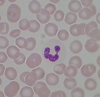
This is a neutrophil.  

  • It has a multilobed nucleus, usually three but it can have more.  
  • Neutrophils phagocytize bacteria, meaning they eat and bust up bacteria that isn't supposed to be there.  
  • It is very difficult to discern granules in a neutrophil because they don't pick up stain well.
  • Neutrophils are just slightly larger than the surrounding red blood cells.
  • Easily the most abundant granulocyte in the blood.


1000x Magnification
This is an eiosinophil.  

  • Still about the same size as a neutrophil.  
  • This guy has a bilobed nucleus and granules that stain RED, not purple.
  • Much less of these than the neutrophil, and harder to find on a slide.
  • Eiosinophils kill parasitic worms and also have some more complicated roles.
Okay so this next one is not one that is easy to find.  They are in 1% of 1% of your blood.  I couldn't find one on any of the slides I looked at. SO.....TO THE INTERWEBS!

Probably more than 1000x?
This is a basophil.
  • Still around the same size as the previous two
  • Bilobed nucleus with big granules that stain dark PURPLE.
  • These guys release histamine, and other inflammation chemicals and also contain heparin


Next time we do agranulocytes.


Send questions or comments to dudaday@gmail.com

Disclaimer:  I am not a health care provider, any information presented in this blog should not be considered advice it is mearly an outlet to slake my curiosity.  You should always consult your primary medical provider for any concerns or illness.  Unlike Tylenol, I am not approved by the FDA or American Medical Association to treat or provide relief for any ailment.

Wednesday, February 13, 2013

Hematopoiesis


Today's Medical Topic:  Hematopoiesis

Today we are looking at the creation or synthesis of blood cells, also known as hematopoiesis.  For the sake of my A&P class and the fact that I have no intention of becoming a hematologist we are simplifying the process and skipping one or more tertiary stages of the genesis.

(click for big, open in new window/tab for original size)
KEY:
IL = interleukin
GM = granulocyte/monocyte
CSF = colony stimulating factor
EPO = erythropoietin

So we have eight final products that we are interested in.  All of them are evolving from the the pluripotent hematopoietic stem cell which is a differentiated stem cell.  This cell may also be referred to as a "self renewal" cell.  

All cells differentiate based on the interleukins and/or granulocyte/monocyte colony-stimulating factors introduced to the precursor cells.

As you can see from the diagram the two major precursor cells are the lymphoid stem cell from which the lymphocytes are generated and the myeloid stem cell from which granulocytes. monocytes, and erythrocytes are generated.

Memorize that diagram for the quiz on Monday and you'll be sound as a pound.


Send questions or comments to dudaday@gmail.com

Disclaimer:  I am not a health care provider, any information presented in this blog should not be considered advice it is mearly an outlet to slake my curiosity.  You should always consult your primary medical provider for any concerns or illness.  Unlike Tylenol, I am not approved by the FDA or American Medical Association to treat or provide relief for any ailment.

Monday, February 4, 2013

Gross Anatomy of the Female Reproductive System


Today's Medical Topic:  Gross anatomy is body parts, structures, and landmarks visible to the naked eye without the aid of a microscope.  Today we are looking at the gross anatomy of the female reproductive system.

What Are We Looking For:  The most obvious parts, most of which are directly related to the reproductive system.  We are looking at a photograph of a model of a mid-sagittal section of the pelvis including part of the abdominal cavity.

If you are in my A&P II class this semester it is the same model in the classroom.

(click for big)
A lot of this is sorta self explanatory but there is some stuff missing as well as some bizarrely presented  areas.

The pubic symphysis is also presented as an almost identical blue oval in this model so I did not point it out specifically.

Three layers compose the wall of the uterus.  The inner deppest layer is the endometrium.  The middle layer, composed mostly of smooth muscle, is the myometrium.  The outer most superficial layer is the perimetrium.

The cervix is a structure that includes the external os, the internal os, and the cervical canal.  The area pointed to labeled "cervix" is specifically pointing to the cervical canal.  The os pointed to is the external os.  Fun fact "os" is Latin for "mouth."

I am also assuming that you know that the urethra leads to the bladder.

Well I didn't Identify everything but most of the stuff.  Don't forget to study!


Send questions or comments to dudaday@gmail.com

Disclaimer:  I am not a health care provider, any information presented in this blog should not be considered advice it is mearly an outlet to slake my curiosity.  You should always consult your primary medical provider for any concerns or illness.  Unlike Tylenol, I am not approved by the FDA or American Medical Association to treat or provide relief for any ailment.

Saturday, February 2, 2013

Gross Anatomy of the Male Reproductive System


Today's Medical Topic:  Gross anatomy is body parts, structures, and landmarks visible to the naked eye without the aid of a microscope.  Today we are looking at the gross anatomy of the male reproductive system.

What Are We Looking For:  The most obvious parts, most of which are directly related to the reproductive system.  We are looking at a photograph of a model of a mid-sagittal section of the pelvis including part of the abdominal cavity.

If you are in my A&P II class this semester it is the same model in the classroom.

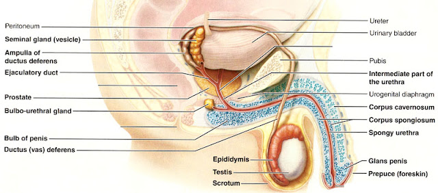
Also we are looking at a picture out of the textbook because some of the structures are not identified or featured on the model.


A lot of this is sorta self explanatory but there is some stuff missing as well as some bizarrely presented  areas.

The pubis is colored blue, I have no idea why since it is bone.  Correction this is the pubic symphysis.  A cartilaginous joint uniting the superior rami of the right and left pubic bones.

There are two parts (technically three but only two are visible in a sagittal section) to the erectile tissue in the penis.  The corpus spongiosum which surrounds the urethra, and the paired dorsal erectile bodies, the corpus cavernosa.  Remember that the anatomy of the penis is named in reference to an erect penis.  So imagine that guy sticking straight out the top part is dorsal, the bottom part ventral.

Not seen on this model is the testes, vas deferens, seminal glands, etc.  Why this is not on the model I have no idea.  This is what we need the picture in the anatomy book for:

(click for big)
I took out some of the labels so it is easier to understand but otherwise it identical to the one in your A&P book.

Fun stuff.


Send questions or comments to dudaday@gmail.com

Disclaimer:  I am not a health care provider, any information presented in this blog should not be considered advice it is mearly an outlet to slake my curiosity.  You should always consult your primary medical provider for any concerns or illness.  Unlike Tylenol, I am not approved by the FDA or American Medical Association to treat or provide relief for any ailment.

Wednesday, January 30, 2013

Ovarian Follicle


We are doing some more histology.  I hope those of you that do not understand the more technical details enjoy the pictures I've taken.

Today's Medical Topic:  Histology of The Ovarian Follicle

What Are We Looking For:  We're looking for oocytes, thecal cells, and granulosa cells among other histological landmarks.

The Tissue Sample:  Alrighty.  Let's take a look.

Particularly we are looking at an antral follicle.  That is, we are looking at an ovarian follicle in the critical preovulatory stage.   Basically, this follicle is about to spit out an oocyte.

So here is the what we are looking at:

(click for big)
Here we can identify a bunch of things:

Thecal cells:  These cells are identified by their wavy appearance.  These cells synthesize androgens which are sex hormones.

Oocyte:  This is the precursor to the "egg."  It remains suspended in prophase I until it is released during ovulation where it undergoes meiosis I in response to a lutenizing hormone surge.

Zona pellucida:  The thin, dark ring around the oocyte.  This is just a glycoprotein membrane surrounding the plasma membrane of an oocyte.  This is the structure that binds to incoming spern during fertilization.

Granulosa cells:  These are spotted/roundish cells that occur around the zona pellucida and the antrum.  These cells synthesize estrogen.

Antrum:  The large mostly empty space between the granulosa cell layers.  This area contains a nutritive liquor.

That was fun, right?  Histology is always awesome.


Send questions or comments to dudaday@gmail.com

Disclaimer:  I am not a health care provider, any information presented in this blog should not be considered advice it is mearly an outlet to slake my curiosity.  You should always consult your primary medical provider for any concerns or illness.  Unlike Tylenol, I am not approved by the FDA or American Medical Association to treat or provide relief for any ailment.

Sunday, January 27, 2013

Histology of the Pancreas


We are doing some more histology.  I hope those of you that do not understand the more technical details enjoy the pictures I've taken.

Today's Medical Topic:  Histology of The Pancreas

What Are We Looking For:  Acinar cells and Islets of Langerhans.

The Tissue Sample:  Alrighty.  Let's take a look.

Just so we are clear here, the pancreas is that tadpole-shaped organ just posterior to your stomach.  The pancreas is a mixed gland composed of endocrine and exocrine glands.  Acinar cells make up most of the glands with Panceatic Islets or Islets of Langerhans sprinkled throughout.

So here is the what we are looking at:

100x Magnification (click for big)
So this is a cross section of part of the pancreas.  What you seeing is mostly acinar cells.  That little red line is pointing to an Islet of Langerhans.  We need to zoom in to really appreciate it.

(click for big)
That blurry little blob there in the middle is an Islet of Langerhans.  These islets contain two major populations of cells:

alpha cells:  These synthesize glucagon

beta cells:  These are much more numerous and synthesize insulin.

I am not able to identify which is which and it is my understanding that alpha and beta cells are very difficult to discern between.  Also it might not be possible with the dye used to stain this sample.

Insulin is needed for your cells to utilize glucose.  Which in turn is needed for ATP production.  And we all know that adenosine triphosphate is your body's energy currency.

I know this was super exciting.  Keep on keepin on and I'll keep on posting.


Send questions or comments to dudaday@gmail.com

Disclaimer:  I am not a health care provider, any information presented in this blog should not be considered advice it is mearly an outlet to slake my curiosity.  You should always consult your primary medical provider for any concerns or illness.  Unlike Tylenol, I am not approved by the FDA or American Medical Association to treat or provide relief for any ailment.

Saturday, January 26, 2013

Histology of the Adrenal Gland


We are doing some more histology.  I hope those of you that do not understand the more technical details enjoy the pictures I've taken.

Today's Medical Topic:  Histology of The Adrenal Gland

What Are We Looking For:  The capsule that encloses the adrenal gland, the three major zones of the adrenal cortex, and the adrenal medulla.

The Tissue Sample:  Alrighty.  Let's take a look.

Adrenal Section at 100x.  (Click for big.)
So here is the whole section we are looking at.  For reference the bottom is the capsule and it goes up through all three layers of the adrenal cortex with the very top of this image being part of the adrenal medulla.

All the layers.  (click for big)
Here I have cut the image in half and identified the areas we are interested in.  From bottom to top:

The Capsule:  The adrenal glands are enclosed in a capsule of fibrous connective tissue.  This is turn is enclosed with a fatty cushion for protection.  This fatty cushion has been mostly dissected away for this slide and is not identified.

The next three sections compose the adrenal cortex:

Zona Glomerulosa:  The thinnest and most difficult to identify part of the adrenal cortex.  Only 5 to 6 cell layers thick this section looks indistinct and blurry.  I have magnified this section below.  This cells of this layer produce mineralcorticoids.

Zona Fasciculata:  The middle layer of the adrenal cortex and the thickest.  Microscopically it looks like cords of cells arranged linearly.  The cells in this layer produce metabolic hormones called glucocorticoids.

Zona Reticularis:  The inner most layer of the adrenal cortex.  Microscopically it is identifiable as a change from cords of lined up cells to a almost gravel-like appearance.  The boarder between this layer and the zona fasciculata are very indistinct but the change in overall appearance is unmistakable.  This layer produces adrenal sex hormones called gonadocorticoids.

Now the inner most section of the adrenal gland.

The Adrenal Medulla:  In the above image you can see about half of the adrenal medulla.  You can see it is quite a large section and also very different from any of the other areas.  The adrenal medulla synthesizes epinephrine and norepinephrine and acts as part of the sympathetic nervous system.

Because it is difficult to see the fibrous capsules and the zona glomerulosa in the above images here is a close up:

(click for big)
I have outlined part of the zona glomerulosa in red to highlight how it is sort of squares of cells.  You can also see the capsule and how the capsule becomes denser as it approaches the adrenal cortex.  I hope this helps those of you studying the endocrine system.  If you have any questions or comments then drop me a line at dudaday@gmail.com.


Send questions or comments to dudaday@gmail.com

Disclaimer:  I am not a health care provider, any information presented in this blog should not be considered advice it is mearly an outlet to slake my curiosity.  You should always consult your primary medical provider for any concerns or illness.  Unlike Tylenol, I am not approved by the FDA or American Medical Association to treat or provide relief for any ailment.