Friday, February 15, 2013

Part Deux of Blood Histology


Today's Medical Topic:  Histology of Blood Part Deux.

What Are We Looking For:  Identifying features, and what makes them what they are.

The Tissue Sample:  Agranulocytes.  If you guessed that agranulocytes have no granules in them then you are a genius, sir and/or madam, and you have no business reading this blog.

Agranulocytes all arise from ancestral stem cells, however, unlike granulocytes, agranulocytes do not all come from a myeloid stem cell.  A monocyte arises from a myeloid stem cell and lymphocytes arise from a lymphoid stem cell.  Lets take a look.

1000x magnification
This is a monocyte

  • Has a kidney-shaped nucleus with an "invagination" which is what the red arrow is pointing to.
  • Is more than three times larger than a red blood cell.
  • Monocytes are the bad boys of the cell world they develop into macrophages and eat stuff.
  • Macrophages can engulf and break down almost anything they can fit inside them.

1000x magnification

This is a lymphocyte

  • Has a spherical nucleus.
  • Is about the same size as a red blood cell
  • Comes in several flavors but lymphocytes mount immune response by direct cell attack or by synthesizing antibodies.

Next up...something.  Something else about blood.


Send questions or comments to dudaday@gmail.com

Disclaimer:  I am not a health care provider, any information presented in this blog should not be considered advice it is mearly an outlet to slake my curiosity.  You should always consult your primary medical provider for any concerns or illness.  Unlike Tylenol, I am not approved by the FDA or American Medical Association to treat or provide relief for any ailment.

Thursday, February 14, 2013

Histology of the Blood Part 1


Today's Medical Topic:  Histology of Blood.  Since we did hematopoiesis it makes sense we should look at the cell's generated in that process one by one.

What Are We Looking For:  Identifying features, and what makes them what they are.

The Tissue Sample:  We have a bunch of samples and we are going to do this in a couple, maybe three parts.  First up granulocytes.

What are granulocytes?  Well they all arise from ancestral stem cells, more specifically, A myeloid stem cell.  They are all characterized by the granules in their cytoplasm that pick up dye.  There are three types.  Eoisinophils, basophils, and neutrophils.

1000x Magnification
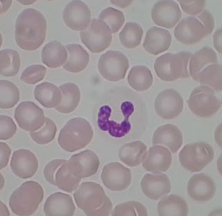
This is a neutrophil.  

  • It has a multilobed nucleus, usually three but it can have more.  
  • Neutrophils phagocytize bacteria, meaning they eat and bust up bacteria that isn't supposed to be there.  
  • It is very difficult to discern granules in a neutrophil because they don't pick up stain well.
  • Neutrophils are just slightly larger than the surrounding red blood cells.
  • Easily the most abundant granulocyte in the blood.


1000x Magnification
This is an eiosinophil.  

  • Still about the same size as a neutrophil.  
  • This guy has a bilobed nucleus and granules that stain RED, not purple.
  • Much less of these than the neutrophil, and harder to find on a slide.
  • Eiosinophils kill parasitic worms and also have some more complicated roles.
Okay so this next one is not one that is easy to find.  They are in 1% of 1% of your blood.  I couldn't find one on any of the slides I looked at. SO.....TO THE INTERWEBS!

Probably more than 1000x?
This is a basophil.
  • Still around the same size as the previous two
  • Bilobed nucleus with big granules that stain dark PURPLE.
  • These guys release histamine, and other inflammation chemicals and also contain heparin


Next time we do agranulocytes.


Send questions or comments to dudaday@gmail.com

Disclaimer:  I am not a health care provider, any information presented in this blog should not be considered advice it is mearly an outlet to slake my curiosity.  You should always consult your primary medical provider for any concerns or illness.  Unlike Tylenol, I am not approved by the FDA or American Medical Association to treat or provide relief for any ailment.
Wednesday, February 13, 2013

Hematopoiesis


Today's Medical Topic:  Hematopoiesis

Today we are looking at the creation or synthesis of blood cells, also known as hematopoiesis.  For the sake of my A&P class and the fact that I have no intention of becoming a hematologist we are simplifying the process and skipping one or more tertiary stages of the genesis.

(click for big, open in new window/tab for original size)
KEY:
IL = interleukin
GM = granulocyte/monocyte
CSF = colony stimulating factor
EPO = erythropoietin

So we have eight final products that we are interested in.  All of them are evolving from the the pluripotent hematopoietic stem cell which is a differentiated stem cell.  This cell may also be referred to as a "self renewal" cell.  

All cells differentiate based on the interleukins and/or granulocyte/monocyte colony-stimulating factors introduced to the precursor cells.

As you can see from the diagram the two major precursor cells are the lymphoid stem cell from which the lymphocytes are generated and the myeloid stem cell from which granulocytes. monocytes, and erythrocytes are generated.

Memorize that diagram for the quiz on Monday and you'll be sound as a pound.


Send questions or comments to dudaday@gmail.com

Disclaimer:  I am not a health care provider, any information presented in this blog should not be considered advice it is mearly an outlet to slake my curiosity.  You should always consult your primary medical provider for any concerns or illness.  Unlike Tylenol, I am not approved by the FDA or American Medical Association to treat or provide relief for any ailment.

Monday, February 4, 2013

Gross Anatomy of the Female Reproductive System


Today's Medical Topic:  Gross anatomy is body parts, structures, and landmarks visible to the naked eye without the aid of a microscope.  Today we are looking at the gross anatomy of the female reproductive system.

What Are We Looking For:  The most obvious parts, most of which are directly related to the reproductive system.  We are looking at a photograph of a model of a mid-sagittal section of the pelvis including part of the abdominal cavity.

If you are in my A&P II class this semester it is the same model in the classroom.

(click for big)
A lot of this is sorta self explanatory but there is some stuff missing as well as some bizarrely presented  areas.

The pubic symphysis is also presented as an almost identical blue oval in this model so I did not point it out specifically.

Three layers compose the wall of the uterus.  The inner deppest layer is the endometrium.  The middle layer, composed mostly of smooth muscle, is the myometrium.  The outer most superficial layer is the perimetrium.

The cervix is a structure that includes the external os, the internal os, and the cervical canal.  The area pointed to labeled "cervix" is specifically pointing to the cervical canal.  The os pointed to is the external os.  Fun fact "os" is Latin for "mouth."

I am also assuming that you know that the urethra leads to the bladder.

Well I didn't Identify everything but most of the stuff.  Don't forget to study!


Send questions or comments to dudaday@gmail.com

Disclaimer:  I am not a health care provider, any information presented in this blog should not be considered advice it is mearly an outlet to slake my curiosity.  You should always consult your primary medical provider for any concerns or illness.  Unlike Tylenol, I am not approved by the FDA or American Medical Association to treat or provide relief for any ailment.

Saturday, February 2, 2013

Gross Anatomy of the Male Reproductive System


Today's Medical Topic:  Gross anatomy is body parts, structures, and landmarks visible to the naked eye without the aid of a microscope.  Today we are looking at the gross anatomy of the male reproductive system.

What Are We Looking For:  The most obvious parts, most of which are directly related to the reproductive system.  We are looking at a photograph of a model of a mid-sagittal section of the pelvis including part of the abdominal cavity.

If you are in my A&P II class this semester it is the same model in the classroom.

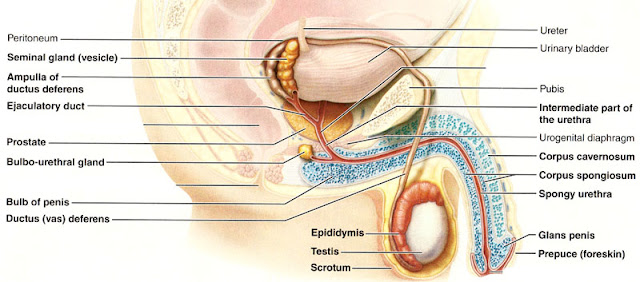
Also we are looking at a picture out of the textbook because some of the structures are not identified or featured on the model.


A lot of this is sorta self explanatory but there is some stuff missing as well as some bizarrely presented  areas.

The pubis is colored blue, I have no idea why since it is bone.  Correction this is the pubic symphysis.  A cartilaginous joint uniting the superior rami of the right and left pubic bones.

There are two parts (technically three but only two are visible in a sagittal section) to the erectile tissue in the penis.  The corpus spongiosum which surrounds the urethra, and the paired dorsal erectile bodies, the corpus cavernosa.  Remember that the anatomy of the penis is named in reference to an erect penis.  So imagine that guy sticking straight out the top part is dorsal, the bottom part ventral.

Not seen on this model is the testes, vas deferens, seminal glands, etc.  Why this is not on the model I have no idea.  This is what we need the picture in the anatomy book for:

(click for big)
I took out some of the labels so it is easier to understand but otherwise it identical to the one in your A&P book.

Fun stuff.


Send questions or comments to dudaday@gmail.com

Disclaimer:  I am not a health care provider, any information presented in this blog should not be considered advice it is mearly an outlet to slake my curiosity.  You should always consult your primary medical provider for any concerns or illness.  Unlike Tylenol, I am not approved by the FDA or American Medical Association to treat or provide relief for any ailment.