Saturday, November 17, 2012

Histology of Mammal Lung


Histology Continued.  Histology is the microscopic study of human tissue.  We are going to be looking at and discussing different tissue types and layers on a microscopic level.  Even if you don't understand it, I think you will at least find some of the pictures I have taken interesting.

Today's Medical Topic:  Histology of Mammal Lung

What Are We Looking For:  I feel like I shouldn't have to explain what a lung is.  But let's first discuss where exactly this piece of tissue is from in the lung.  My buest gess is that is is a transverse section that includes a small branch of trachea.


So obviously the slide cannot hold an entire transverse section of the lung but definitely part of it.

The Tissue Sample:  So this is mammal lung.  What can we expect to see?  For one we can expect some sort of a piece of trachea or a trachea branch and all the associated tissues with that.  We are going to see part of some alveoli air sacs.  And we are probably going to see some sort of connective tissue.

40x Magnification
The squiggly stuff is alveoli.  Alveoli is composed of simple squamous epithelium.  Ideal for fast filtering but only one cell layer thick.  It is extremely distinctive.  No other tissue type looks quite like it.

The next thing down labeled "This thing" looks like some sort of tracheal branch.  We are going to look at that close up and discuss it in detail.  The next part down I sincerely have no idea what that is supposed to be. I think it is just some degraded tissue because the slide is old.  At any rate we will ignore it for the sake of simplicity.

250x Magnification
So here is a close up of the tracheal branch called a bronchiole.  You can see it composed of more than one distinctive tissue types.  You can see some smooth muscle there.

The darker pink/purple "chunks" that you see are the inner lining of the bronchiole.  This should be either cuboidal or columnar epithelia depending on the size of the bronchiloe.  In this case it appears to be ciliated columnar epithelial tissue.  The ciliated part is for moving dust and debris particles out of the lung.  Lets get closer.

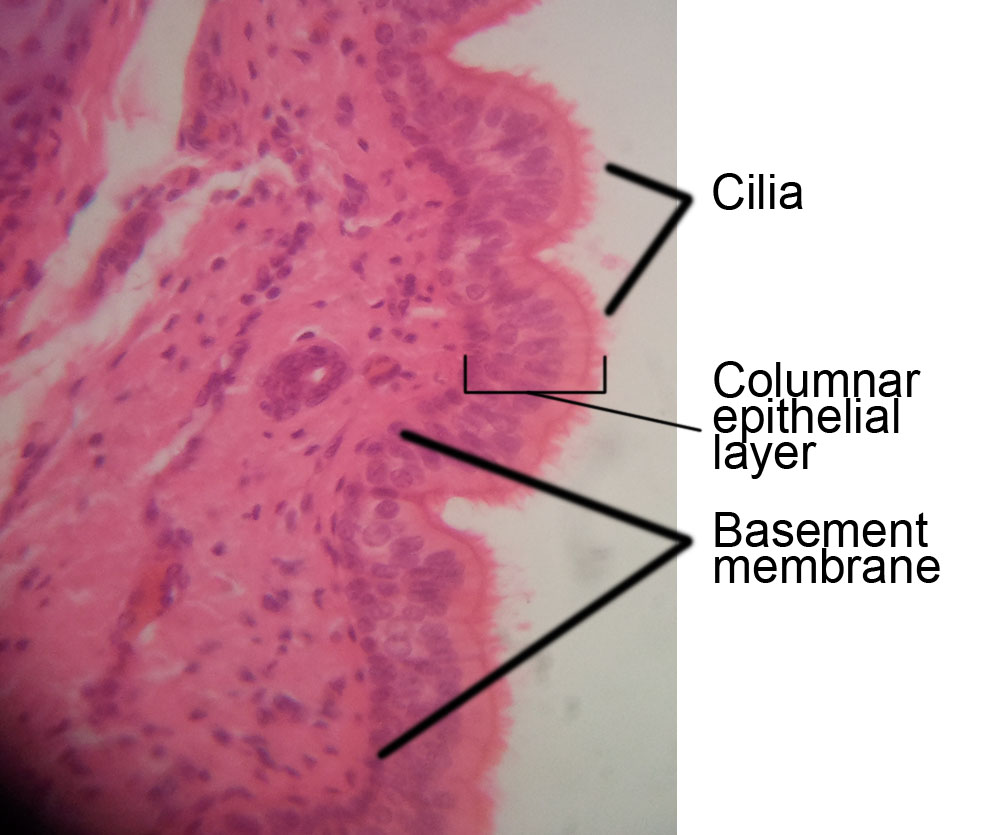
400x Magnification
Here you can clearly distinguish the cilia and also the darker pink line that is the basement membrane of the columnar epithelial layer.

The white space in the middle is just that.  Empty space.  That is the passageway in which air moves when you inhale and exhale.  There are some other tissues scattered about that are difficult to identify with whatever stain was used to dye this slide.

We can assume there is probably some sort of connective tissue in there and also some other passageways and structures which are beyond the scope of my knowledge.  But there is definitively those three distinct tissue types as mentioned above with all of their inherent structures.

Well, this post took way too long.  Next we will look at something simpler.


Send questions or comments to dudaday@gmail.com

Disclaimer:  I am not a health care provider, any information presented in this blog should not be considered advice it is mearly an outlet to slake my curiosity.  You should always consult your primary medical provider for any concerns or illness.  Unlike Tylenol, I am not approved by the FDA or American Medical Association to treat or provide relief for any ailment.

Sunday, November 11, 2012

Epiglottis Histology


We are going to be doin histology for a while.  Histology is the microscopic study of human tissue.  We are going to be looking at and discussing different tissue types and layers on a microscopic level.  Even if you don't understand it, I think you will at least find some of the pictures I have taken interesting.

Today's Medical Topic:  Histology of the Epiglottis.

What Are We Looking For:  The epiglottis is a little flap of cartilage that folds over your trachea when you swallow so you don't inhale food and water.  The epiglottis is constructed of a specific type of cartilage called elastic cartilage.  This type of cartilage is unique and really only found in a few places in the body, namely your ears and the epiglottis.

So what are we looking for?  Well first off we want to find the section of elastic cartilag this section should be very distinctive.  We will then discuss the surrounding tissue and how to identify that tissue microscopically.  Are you ready?  Let's do this.

The Tissue Sample:  This is a slide of a section of a human epiglottis and it's surrounding tissue.  It is dyed specifically to show off the elastic cartilage.  So probably the thing that is the darkest, or has soaked up the most dye, is probably the epiglottis.

40x Magnification
So the purple stuff is probably elastic cartilage.  Which is correct.  That is what it is.  As you can see the epiglottis is not just  composed of elastic cartilage, it has other tissues as well.  Let's zoom in.

250x Magnification (Click to make bigger)
Here the elastic cartilage is becoming more distinctive.  Looking closer you can see a thin tissue layer that boarders the cartilage.  This is probably dense irregular connective tissue.  And the larger, lighter areas of wispy tissue adjacent to that is probably loose adipose connective tissue.  It is interspersed with sections of more dense irregular connective tissue.  Let's get closer to see how distinctive these layers are.

400x Magnification
Here we can see how very distinctive everything is.  The elastic cartilage is unmistakable.  Can you see how the fibers in the dense irregular tissue goes in all different directions and are very close together?  And how the adipose tissue shows big globules of fat that take up most of the cell volume?

These distinguishing features do not change between slides.  They might be different colors, but don't let that shake you.  You might even see multiple colors in slides to highlight different features but the shapes and arrangements will stay the same.

Keep it tuned here for more histology.  Tomorrow we look at mammal lung.


Send questions or comments to dudaday@gmail.com

Disclaimer:  I am not a health care provider, any information presented in this blog should not be considered advice it is mearly an outlet to slake my curiosity.  You should always consult your primary medical provider for any concerns or illness.  Unlike Tylenol, I am not approved by the FDA or American Medical Association to treat or provide relief for any ailment.

Saturday, November 10, 2012

Saxitoxin


This is the final part of my three part series on neurotoxins.

Today's Medical Topic:  Saxitoxin.

Off the Top of My Head:   I honestly know nothing about this.

My Research Today:  Saxitoxin is associated with a condition known as Paralytic Shellfish Poisoning. It is most commonly introduced to humans when they consume shellfish that is contaminated with the toxin.  The shellfish or mollusks must filter feed a specific kind of dinoflagellate algae to become contaminated and this algae is what produces this very potent neurotoxin.

Saxitoxin is thought to be one of the most toxic naturally occurring non-protein substances in the world.  A dose as small as 0.2mg can be fatal to humans.  To put that into perspective a standard dose of Tylenol is 650mg.  Luckily for us most times we do not consume that much toxin and Paralytic Shellfish Poisoning is rarely fatal.

Saxitoxin, apparently.
Symptoms include tingling, numbness, weakness and paralysis which usually manifest within 12 hours if you have ingested the poison.  If all else fails you can place a patient on life support until they metabolize the poison.  Your body will breakdown damn near anything if you can keep it alive long enough.

So how does Saxitoxin work?  Once digested Saxitoxin finds itself into your blood stream diffusing throughout your body.  It selectively binds to voltage-gated sodium channels closing them and blocking passage of the nerve impulse.  If you can't move sodium ions, you can't change your membrane potential.


The difference between this toxin and curare is that this one doesn't compete with acytelcholine it just jams itself into the ion channel.  ACh can bind if it wants but it won't do dick because the channel is blocked with a big ass molecule of saxitoxin.

So there you go.  I hope you enjoyed this series.  Tomorrow we start on histology!


Send questions or comments to dudaday@gmail.com

Disclaimer:  I am not a health care provider, any information presented in this blog should not be considered advice it is mearly an outlet to slake my curiosity.  You should always consult your primary medical provider for any concerns or illness.  Unlike Tylenol, I am not approved by the FDA or American Medical Association to treat or provide relief for any ailment.

Friday, November 9, 2012

Curare


We are continuing my 3 part series on neurotoxins.  Today we are discussing Curare.  Everyone needs to put on their big girl pants for this one because it gets technical.  All the way technical, which is the best kind of technical.

Today's Medical Topic:  Curare:  What is it, how does it work, and why is my tongue numb?

Off the Top of My Head:   An almost mythical poison the actuality of what curare is has become quite skewed throughout history.  In novels like Sherlock Holmes it is the mysterious poison created by witch doctors in jungle tribes and used with devastating accuracy in blow guns.  But how much of this is true and how much is myth?  Let's find out.

My Research Today:  "Curare" used by South American tribes is actually a strange syrupy concoction of plants, roots, toxins, and venoms assembled from various sources.  These items are crushed, cooked, and mixed to make a black sticky poison that tribal hunters use on the tips of their arrows.

They aren't wearing much but I feel
like 90% of it is still frivolous.
Regardless of what else is mixed into this concoction the active ingredient of curare is D-tubocurarine the product of a root of a vine called Chondroendron tomentosum.  D-tubocurarine is a non-depolarizing muscle relaxant drug (NDMRD).  Many NDMRD's have been developed from D-tubocurarine but let's look at how this class of drug works.

The name gives you a hint as to how it works, what with the non-depolarizing in the title, but if you look closer you will see it is more complicated than that.  NDRMD's diffuse through blood vessels into the synaptic cleft of motor neuromuscular junctions.  There NDMRD's compete with acetylcholine (ACh) molecules released into the synaptic cleft and then bind to the ACh receptors on the synaptic endplate.  This blocks the ACh from binding and therefore prevents the initiation of the depolarization process of muscle contraction.


NDMRD's also act on presynaptic receptors which interfere with the entry of calcium ions into the axon terminal which in turn doesn't allow as much ACh to be released magnifying the effect of the NDRMD.

It is importanant to note the NDMRD's must be injected into the bloodstream and cannot work if they are simply ingested by other means.  NDMRD's follow a simillar paralyzation pattern as Botulism does. Starting from top to bottom.  You need a fairly significant dose for NDMRD's to affect your chest wall muscles and diaphragm.

NDMRD's are a water-soluble, polar molecules which make them unable to break the blood-brain barrier.  For this reson NDMRD's do not effect your autonomous nervous system so your heart will continue to beat.  This makes NDMRD's ideal for sedated patients on ventilators.

That is all for now.  Tomorrow we work on Saxitoxin which is somewhat difficult to find data on..


Send questions or comments to dudaday@gmail.com

Disclaimer:  I am not a health care provider, any information presented in this blog should not be considered advice it is mearly an outlet to slake my curiosity.  You should always consult your primary medical provider for any concerns or illness.  Unlike Tylenol, I am not approved by the FDA or American Medical Association to treat or provide relief for any ailment.

Thursday, November 8, 2012

Botulism


I'm back at it.  Sorry for my absence.  I have been studying hard in school but now I am bringing school to you in a three part series on neurotoxins.  One of my favorite topics.  Gotta love poisons.

Today's Medical Topic:  First up an age old favorite:  Botulism.

Off the Top of My Head:   The scourge of people who improperly can food and kill their relatives; botulism is an anaerobic bacteria.  That means it can grow in an oxygen-free liquid environments like canned food or raw honey.

Most people these days know of botulism in it's derived form of Botox, the custom poison that people inject in their face to loosen wrinkles. Botox keeps all the paralytic fun of botulism without the massive infection that kills you.

My Research Today:  Botulism works by hijacking your motor axon terminals at the neuromuscular junction.  It tricks the axon terminal membrane to let it in through endocytosis and now the botulism bacteria is contained in it's own vesicle in the axon cell.

Once inside the botulism ejects part of it's self out of it's vesicle into the cytoplasm of the axon terminal.  This part directly interferes with vesicles of acetylcholine (ACh) that is bound for the synaptic cleft and traps it right before it can deliver it's payload.  No acetylcholine, no muscle contraction.

Here is a clumsy drawing of sort of how it works.
Botulism also dumps itself into the synaptic cleft where it continues to replicate and spread.

Neurologically botulism effects you from the top down.  It starts with your cranial nerves cause paralysis of the face and continuing down the body bilaterally.  Eventually it will paralyze your chest wall muscles and diaphragm making it impossible for you to breathe.  You can get other nice symptoms like nausea and vomiting prior to the paralysis.

Here is what makes botulism extra evil though:  It doesn't effect your sensory axons or your mental status.  So you are fully conscious and able to feel everything but unable to move, speak, or do anything about your impending doom.

Keep it tuned here for more information.  Tomorrow we do Curare.  Exciting.


Send questions or comments to dudaday@gmail.com

Disclaimer:  I am not a health care provider, any information presented in this blog should not be considered advice it is mearly an outlet to slake my curiosity.  You should always consult your primary medical provider for any concerns or illness.  Unlike Tylenol, I am not approved by the FDA or American Medical Association to treat or provide relief for any ailment.

Saturday, September 1, 2012

Mitochondria


**NOTE**  Sorry I have not posted recently, most of my time has been taken up by school.  Over the Labor Day weekend I have a little extra time so I am going to be posting as much as possible.

Today's Medical Topic:  Mitochondria

Off the Top of My Head:   Often called :the power plant of the cell" mitochondria is an organelle within cells that somehow provide or generate energy.  How do they do that?  I am not entirely sure but I am going to try to find out.

Sorta looks like the lazy river in a water park.


My Research Today:  Mitochondria are organelles located in the cytoplasm of your cells.  Mitochondria are fairly large, respectively, and can be seen on a 10,000x microscope with an oil immersion lens.  There are multiple mitochondria per cell and they are responsible for manufacturing a special molecule that provides energy for your cells.

Primarily the function of mitochondria is cellular respiration and to produce adenosine triphosphate or ATP which is a high energy fuel source for every cellular reaction in your body.  Mitochondria also does some other stuff too but we are not going to talk about that today.

Damn carbon molecules are like
dog crap.  They get everywhere.
The process starts by cutting one glucose molecule into two smaller glucose molecules.  This process also yields 2 ATP molecules, some sort of acid, and 2 electron carrying molecules.  The two sugars are converted into a different compound, this in turn produces 2 more ATP molecules plus several more compounds that carry electrons.  This cycle is called the "Citric Acid Cycle" and it only occurs when oxygen is present in the hemoglobin, however, it does not use oxygen directly.

I have no idea.
This next step is where your mitochondria comes in.  The mitochondria channel the electron string into the oxygen and through some extremely complex chemical reactions this creates even more ATP.  The cycle repeats itself as needed as long as there is fuel and oxygen to burn.

I hope I did not screw that up too bad.  Maybe tomorrow I will get into what exactly adenosine triphosphate is.  And maybe I will stick with subjects I have a better understanding of.


Send questions or comments to dudaday@gmail.com

Disclaimer:  I am not a health care provider, any information presented in this blog should not be considered advice it is mearly an outlet to slake my curiosity.  You should always consult your primary medical provider for any concerns or illness.  Unlike Tylenol, I am not approved by the FDA or American Medical Association to treat or provide relief for any ailment.

Thursday, August 23, 2012

Septic Shock


Today's Medical Topic:  The spleen.  What does it do?  Why do we have one?  Let's find out.

Off the Top of My Head:   Septic shock is a really terrible outcome of septicemia which is a diffuse infection in your blood stream. I know septic shock is something you rarely survive.  I know that it is a continuous positive feedback loop that creates a cascade event that kills you but I am unsure of the mechanics and chemical changes that occur to make this happen.

Even I understand this feedback loop.


My Research Today:  Sepsis is caused by a bacterial, fungal, or (rarely) viral infection that has gotten into your blood stream.  It is more common in elderly and very young people.  Septic shock is an acute inflammatory response to the infection in your blood stream which can cause all sorts of problems leading to death.

It can also be caused by Snookie
This inflammatory response from the invading pathogen is actually from the toxins the pathogen produces.  This in turn can cause a cascading multiple organ failure in which your body cannot maintain homeostasis due to the organ damage from the toxins.  This will lead to cardiogenic shock which is the inability of your heart to output an adequate amount of oxygenated blood.  This causes cell death throughout your body.

The inflammation can also cause pulmonary edema making your lungs fill with fluid and then you drown in your own juices.  I am not sure if this is preferable to the above option or not.

Pulmonary edema can also be caused by Snookie.
So in this case the inflammatory response is a positive feedback loop of your body overworking to try to fight the infection which ends up making things worse.  As it progresses it compounds the inflammation exponentially.

Most management is supportive care, like a mechanical ventilator, hemologic or support to keep your blood pressure up.  Your body either survives until antibiotics can take effect or dies due to massive organ damage.

Welp, I am done for the day.


Send questions or comments to dudaday@gmail.com

Disclaimer:  I am not a health care provider, any information presented in this blog should not be considered advice it is mearly an outlet to slake my curiosity.  You should always consult your primary medical provider for any concerns or illness.  Unlike Tylenol, I am not approved by the FDA or American Medical Association to treat or provide relief for any ailment.